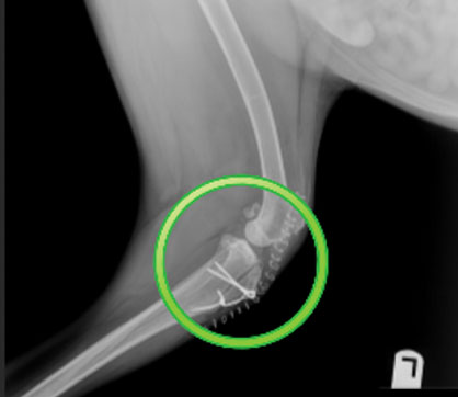
Pet Patellar Luxation
Patellar luxation is a condition in which the patella (kneecap) slips out of its normal location within the stifle (knee). Patellar luxation has been described as the most common congenital, or present at birth, malformation in dogs diagnosed in 7% of puppies and can manifest in different types of luxation and varying grades of severity. Small-breed dogs are affected at 10 times the rate found in large breed dogs.
Pet Patellar Luxation in Toledo, OH
WHAT ARE THE SYMPTOMS OF PATELLAR LUXATION?
The clinical signs of patellar luxation vary greatly with the severity of the disease, ranging from non-symptomatic to complete lameness in the affected leg. Sometimes, signs will come and go as the patella luxates and then moves back into the correct location.
Patellar Luxations are graded according to severity as follows:
- Grade I: Patella is loose but generally stays in the groove. Your dog may occasionally skip/hop, and is painful only when the patella is luxated. Most dogs with Grade 1 patellar luxation are only mildly affected, and surgery is often not needed. Most vets are able to luxate the patella and return it to its normal position.
- Grade II: The patella is in a normal position more than it is out. Frequency of luxation increases, and the dog carries the leg more often. By manipulation, the vet can return the patella to a normal position.
- Grade III: The patella is out more than in. Some dogs may appear bow-legged. By manipulation, the vet can return the patella to a normal position, but it will spontaneously pop back out when the dog moves the leg through its normal range of motion.
- Grade IV: The patella is permanently luxated. The dog may not weight-bear on that leg at all. At this point, the vet cannot return the patella to a normal position by manipulation alone.
Other conditions like a torn ligament inside the knee can also accompany patellar luxation. The doctor will explain what procedure is best for your pet at the time of the consultation.

PATELLAR LUXATION CORRECTION FOR DOGS
If your dog has Grade II, Grade III, or Grade IV patellar luxation, surgery is the only means of correcting the issue. There are four different techniques we can use to correct patellar luxation:
Trochleoplasty – The trochlear groove is located at the end of the femur, right above the knee. In dogs with patellar luxation, this groove is usually shallow. A trochleoplasty is one means of deepening this groove and thereby preventing future luxation of the patella.
Tibial tuberosity transposition – Dogs can either have medial patellar luxation where the patella moves toward the inside of the knee, or lateral patellar luxation, where it moves to the outside. TTT surgery moves the point, where the patella attaches to the tibia toward the outside of the knee for medial luxation and toward the inside for lateral luxation.
Lateral imbrication and medial release – In this surgery, the tissues surrounding the patella are loosened or released on the inside of the knee and tightened (imbricated) on the outside of the knee for medial patellar luxation. The opposite is done for lateral patellar luxation.
Some dogs can also have an abnormal curvature of the lower part of the femur (thigh bone) that contributes to patellar luxation. In more severe cases, a corrective procedure may be needed to straighten the femur in addition to the above techniques to correct the patellar luxation.
To schedule your consultation, call us at 419-841-4745 or make an appointment online.
REFERRAL SURGICAL SERVICES
Have you been referred to West Suburban Animal Hospital for patellar luxation correction? Don’t forget to read the Referral Policy and bring these important things to your consultation:
- Any recent radiographs or blood work from your family veterinarian
- Any current medications your pet is taking
- A completed Referral Form
To schedule your consultation, call us at 419-841-4745 for more information.


